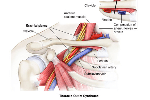
The thoracic outlet is the part of the body surrounded by the curvature of the first rib on each side; this is the base of the neck and the top of the chest. The blood vessels (arteries and veins) and nerves to the arm exit from the chest through this anatomic area, i.e., it is the outlet for these structures which get their start in the thorax. A network of muscles, tendons and ligaments interlace in this tight space and the blood vessels and nerves weave between and among them on the way to the arms. The thoracic outlet syndrome is produced if a major artery, vein or nerve is compressed between two of these structures or against a bone such as the first rib, the clavicle (the collar bone) or an aberrant rib called a cervical rib, found in some people, connected to the spine in the neck rather than the spine in the chest from which the typical rib arises.
The syndrome is well described and understood; however, in practice it is somewhat controversial. Signs and symptoms of nerve compression are most frequent and include tingling, numbness, pain and muscle atrophy. Artery compression causes pain in the arm, especially with activity. Venous compression leads to thrombosis (clotting) of the vein. There are a variety of tests and physical exam findings used to make a diagnosis. There are two challenges. The test and exam results are frequently equivocal and not definitive. Additionally, this diagnosis is occasionally used and abused by people seeking worker compensation.
Treatment begins with physical therapy to strengthen the muscles of the shoulder girdle; patients with neurologic symptoms are the most likely to benefit. When the surgeon is confident of the diagnosis and physical therapy proves insufficient an operative procedure to decompress the contents of the thoracic outlet is appropriate. There are several operations in use; of increasing popularity is using a robotic approach to resect the first rib which decompresses the contents of the thoracic outlet.


